Back to Blog
July 7, 2026
Breast Ultrasound vs. Mammogram: What’s the Difference (and Do You Need Both)?
Mammograms and breast ultrasounds do different jobs. How each works, where each falls short, and why women with dense breasts often benefit from both.
Share on:

If you’ve started looking into your breast screening options, you’ve probably run into a confusing choice: breast ultrasound vs mammogram. Are they the same thing? Is one better? Can you skip the uncomfortable one?
Here’s the short, honest answer up front: they’re different tools that do different jobs, and for many women, especially those with dense breasts, the goal isn’t choosing one instead of the other. It’s understanding how they work together.
Let’s break down the real differences, without the sales pitch, so you can have a well-informed conversation with your provider.
What a mammogram does
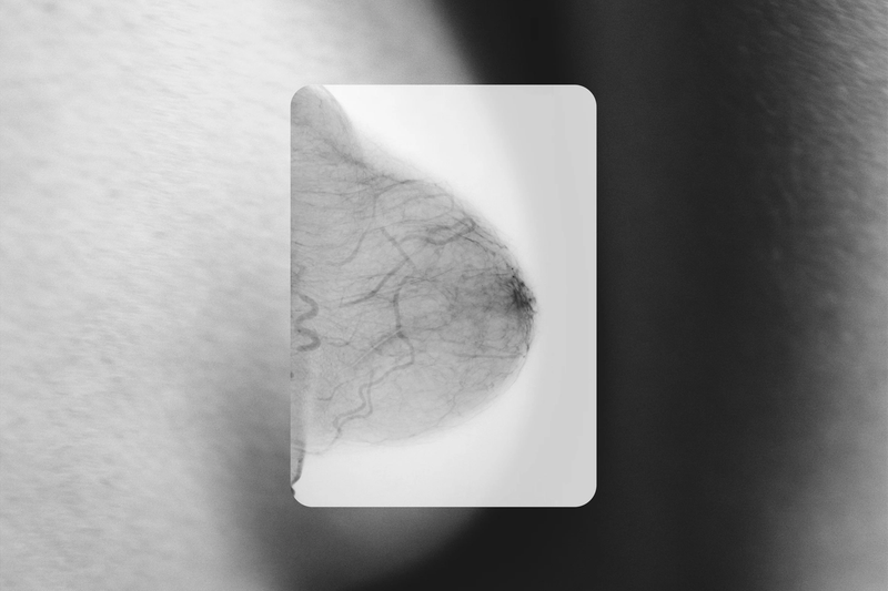
A mammogram is an X-ray of the breast. The breast is compressed between two plates, and low-dose radiation captures a 2D (or, with a 3D mammogram, a layered) image of the tissue.
Mammography is the foundation of breast cancer screening for good reason. It’s the most studied breast imaging tool in existence, it’s excellent at spotting tiny calcium deposits (microcalcifications) that can be an early sign of cancer, and decades of data show it saves lives. If you’re 40 or older and average risk, this is your starting point.
Its main limitation is one of physics: on a mammogram, dense tissue appears white, and so do tumors. In dense breasts, that overlap can hide cancers. It’s not a flaw in the machine; it’s the nature of using X-rays on dense tissue.
What a breast ultrasound does
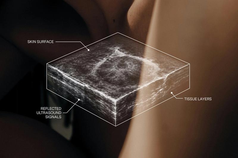
A breast ultrasound uses sound waves, the same safe technology used to image babies during pregnancy, to build a picture of what’s inside the breast. No radiation. No compression. No dye.
Because ultrasound distinguishes tissue by density and texture rather than by X-ray absorption, it can often “see through” dense fibroglandular tissue where a mammogram gets stuck. That’s why the question is ultrasound better for dense breasts comes up so often, and in that specific situation, ultrasound adds something a mammogram can’t. It’s also the go-to tool for taking a closer look at a lump or an area flagged on a mammogram.
Its limitation? Ultrasound is less reliable at catching those tiny microcalcifications that mammograms excel at. Which is the whole point of this article: neither tool is “the best.” They cover each other’s blind spots.
Breast ultrasound vs. mammogram: side by side
- How it works: a mammogram uses low-dose X-ray. Ultrasound uses sound waves.
- Radiation: mammogram, yes (low dose). Ultrasound, none.
- Compression: mammogram, yes. Ultrasound, none.
- Great at: mammogram, microcalcifications and overall screening. Ultrasound, seeing through dense tissue and evaluating lumps.
- Struggles with: mammogram, dense tissue can hide cancer. Ultrasound, less reliable for microcalcifications.
- Best role: mammogram, the foundation of screening (age 40+). Ultrasound, a powerful supplement, especially for dense breasts.
Can I get a breast ultrasound instead of a mammogram?
This is one of the most common questions, and the answer is important: generally, no, not as a replacement. For women at average risk, ultrasound is not considered a stand-alone substitute for a screening mammogram. The two are designed to work as a team.
Where ultrasound shines is as a supplemental layer on top of your mammogram, particularly if you have dense tissue. Think of your mammogram as the wide-angle lens and ultrasound as the tool that fills in what dense tissue obscures. When added to mammography, automated breast ultrasound detects 35.7% more breast cancers than mammography alone (FDA PMA P110006).
If you’re under 40 and not yet eligible for routine mammograms, that’s a different conversation to have with your provider. Ultrasound is sometimes used for proactive screening in younger women, since their breast tissue tends to be denser too.
Is ultrasound or MRI better for dense breasts?
MRI is the most sensitive breast imaging tool available; it catches the most. But it comes with trade-offs: it’s expensive, it requires an injection of contrast dye, it takes much longer, and it’s generally reserved for women at high risk (for example, those with a strong family history or a known genetic mutation).
For the large group of women who have dense breasts but aren’t otherwise high-risk, ultrasound often hits the sweet spot: no radiation, no dye, no compression, widely accessible, and far more affordable, while still seeing well in dense tissue. It’s the thorough middle ground between a mammogram alone and a full MRI.
“What percentage of breast ultrasounds are cancer?”
If you’ve been called back for an ultrasound and you’re anxiously searching what percentage of breast ultrasounds are cancer, here’s some reassurance: the vast majority of breast ultrasounds do not find cancer. Most scans are completely clear or show benign, harmless findings like cysts or fibroadenomas (common non-cancerous lumps).
Being asked to come in for an ultrasound is routine, not a red flag. It usually just means a radiologist wants a clearer look, often precisely because dense tissue made the mammogram harder to read. More looking is a sign of thoroughness, not bad news.
The bottom line
You don’t have to pick a side in the breast ultrasound vs. mammogram debate. For most women:
- Mammogram: your essential foundation.
- Ultrasound: a powerful supplement, especially if you have dense breasts.
- MRI: reserved for higher-risk situations.
The right combination depends on your density, your risk factors, and a conversation with your provider.
Modern supplemental ultrasound in Los Angeles
If you have dense breasts and want that added layer of clarity, BeSound offers advanced automated breast ultrasound in Los Angeles. It’s a comfortable 20-minute scan (no radiation, no compression, no dye), reviewed by a board-certified physician, with results in your secure portal within 24 to 48 weekday hours.
It’s built to do exactly what this article describes: work alongside your mammogram to catch what dense tissue can hide. Same women-first care, no cold waiting rooms, upfront pricing, and answers you can trust.
Curious whether supplemental screening makes sense for you? Book a breast ultrasound in Los Angeles, or take our free breast cancer risk assessment to understand where you stand first.
Not medical advice. Consult a medical professional with any medical questions. BeSound provides supplemental breast ultrasound imaging and does not replace mammography or medical care. Imaging is interpreted by licensed physicians. For marketing purposes only. The American Cancer Society recommends that women at average risk begin regular mammograms at age 40. BeSound is a technology platform that connects individuals with independent partner medical groups that provide breast imaging services; BeSound is not a healthcare provider.
Read more
Start your BeSound journey
Book your scan, understand your lifetime risk, and view your results directly in your BeSound dashboard.
Join the waitlist
Our modern breast imaging experience is coming to more cities across the U.S. Join the waitlist to be the first to know when we launch in your city.
Learn your risk
Our free assessment gives results based on your unique health profile using a clinically validated model trusted by healthcare providers.