Back to Blog
July 7, 2026
What Is ABUS? Automated Breast Ultrasound vs. Traditional Handheld Ultrasound
ABUS and handheld ultrasound sound identical but work very differently. What automated breast ultrasound is, how it compares, and why standardization matters.
Share on:
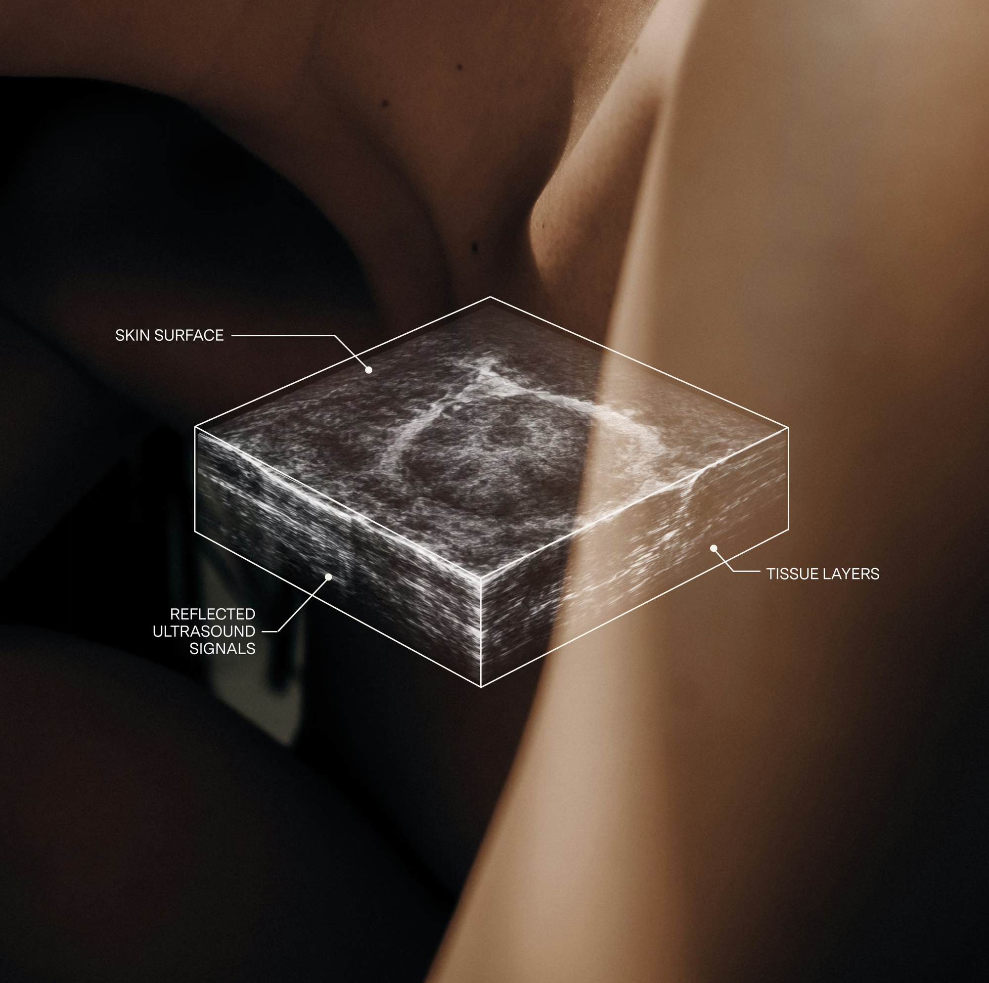
If you’ve been researching supplemental breast screening, you may have run into two terms that sound almost identical: automated breast ultrasound (ABUS) and traditional handheld ultrasound. Both use sound waves, both avoid radiation, and both can see in dense tissue. So it’s easy to assume they’re the same scan. They aren’t.
The differences come down to how the image is captured, how consistent it is, and how much of the breast actually gets seen. If you have dense breasts and you’re weighing your options, here’s what sets ABUS apart.
What ABUS stands for
ABUS stands for Automated Breast Ultrasound System. Like any ultrasound, it uses sound waves, not radiation, to create images of the inside of the breast. The word that matters most is automated, and it’s the whole reason ABUS exists as its own category of screening.
ABUS vs. traditional handheld ultrasound: the core difference
With a traditional handheld ultrasound, a sonographer holds a small probe and moves it around your breast by hand, watching a live 2D image on a screen. Where the probe goes, how long it lingers, and which areas get captured all depend on the person performing the scan. It’s a skilled job, but it is operator-dependent, which means the result can vary from one technician, and one facility, to the next.
An automated breast ultrasound works differently. Instead of a probe guided by hand, ABUS uses a large, curved transducer that sweeps across the whole breast along a set path and captures it as a standardized, high-resolution 3D volume. The same complete view is recorded every time, for every patient, regardless of who runs the machine. Nothing gets skipped because someone was rushing or interpreting the screen in real time.
That single shift, from hand-guided and real-time to automated and standardized, is what drives every other difference below.
- Coverage: ABUS scans the entire breast along a set, automated path. With handheld ultrasound, the operator directs the probe to chosen areas.
- Image: ABUS captures a comprehensive 3D volume, including the coronal view. Handheld ultrasound produces real-time 2D images.
- Consistency: ABUS is standardized and reproducible, easy to compare to priors. Handheld results depend on operator technique.
- Reading: ABUS pairs with AI-assisted interpretation that supports the physician. Handheld ultrasound has no AI-assisted workflow.
- FDA status: ABUS is FDA-approved for supplemental screening in dense breasts. Handheld ultrasound is FDA-cleared for general use.
- Results: ABUS results typically arrive within 24 to 48 weekday hours. Handheld turnaround varies by facility.
Why “standardized and reproducible” matters more than it sounds
Reproducibility isn’t a technical footnote. It’s one of the most practical advantages ABUS has over handheld scanning.
Because ABUS records the whole breast the same way every time, this year’s scan can be lined up against last year’s and compared directly. A radiologist can see whether an area has changed, appeared, or stayed exactly the same. With handheld ultrasound, the images reflect wherever the probe happened to travel that day, so a clean, side-by-side comparison over time is much harder to achieve. For tracking dense tissue year over year, that consistency is a real advantage.
Standardization also supports better consistency and fewer false positives than operator-dependent handheld scanning. A complete, reproducible dataset gives the reader more context to work from.
The coronal view: seeing the whole breast, not just slices
Handheld ultrasound shows the breast in flat, real-time slices as the probe moves. ABUS goes a step further by reconstructing a full 3D volume, which includes a special perspective called the coronal plane, a view that handheld scanning cannot recreate the same way.
Think of it as the difference between flipping through a few loose photographs and having a full model you can rotate and view from any angle. That whole-breast, any-angle view helps a radiologist pinpoint a finding and see how tissue relates across the entire breast, not just at the spot where a probe was placed.
Where does the AI come in?
Modern ABUS is paired with AI-assisted reading. BeSound uses QVCAD, the first FDA PMA-approved AI system for concurrent reading of ABUS exams. As a board-certified physician reviews your images, AI-assisted reading flags areas for closer review, acting as a consistent second set of eyes. Traditional handheld ultrasound has no comparable AI-assisted workflow.
The goal isn’t to replace the physician. It’s to support them, helping flag areas for closer review and keeping the read consistent. A board-certified physician always makes the final assessment on your results.
So is handheld ultrasound still useful?
Yes, and it’s worth being fair about this. Handheld ultrasound is an excellent tool for targeted questions: taking a close, real-time look at a specific lump you or your doctor can feel, zooming in on an area flagged by another scan, or guiding a needle during a biopsy. In those moments, real-time, operator-directed imaging is exactly what you want.
The distinction is screening versus targeted evaluation. ABUS is built to screen the entire breast in a standardized, reproducible way, which is what supplemental screening for dense tissue calls for. Handheld shines once the question has already narrowed to a specific spot. Many women will encounter both at different points, and that’s completely normal.
What does breast cancer look like on an ultrasound?
Patients often ask “what does breast cancer look like on ultrasound”, usually while waiting on results. The honest answer is that interpreting these images takes years of specialized training, and it’s not something to attempt yourself. In broad terms, benign findings like cysts (fluid-filled sacs) tend to look smooth, round, and dark, while areas worth a closer look may appear more irregular. But features overlap, subtlety is everything, and that’s exactly why your scan is read by a board-certified physician.
How long does a breast ultrasound take?
Most people wonder “how long does a breast ultrasound take”, and here ABUS has a practical edge. Because the scan follows a set, automated path, it’s typically about 15 to 20 minutes for both breasts, and the experience stays consistent from visit to visit. A thorough handheld whole-breast ultrasound, by contrast, can be more time-intensive and varies with the operator. From start to finish at BeSound (arriving, changing into a robe, and the scan itself), you’re usually in and out in around 30 minutes.
There’s no compression and no radiation. You’ll feel light pressure and warm gel as the transducer moves across the breast, but nothing painful. And because ABUS uses sound waves with no ionizing radiation, it’s safe for repeated use.
How much does a breast ultrasound cost?
Pricing for breast imaging is often frustratingly opaque, so here’s a straightforward answer. If you’re asking how much does a breast ultrasound cost, the number varies widely by provider, which is exactly why upfront pricing matters. At BeSound, an ABUS scan is $349. You’ll know the cost of your imaging before you book it, not after.
Getting an ABUS scan in Los Angeles
BeSound brings automated breast ultrasound to Los Angeles in a space that feels nothing like a sterile hospital wing. Our flagship location on Melrose Avenue was designed with your comfort in mind: an all-female sonographer team, warm and private rooms, and a calm, unhurried experience.
Here’s the whole journey: you complete your intake and consent online, you can take our free risk assessment, a clinician reviews your information and places the appropriate order, you come in for your 20-minute scan, and a board-certified physician reviews your images. Results land in your secure portal, typically within 24 to 48 weekday hours. If anything needs a closer look, a patient care coordinator walks you through the next steps. You’re never left to figure it out alone.
If you have dense breasts and you want screening that covers the whole breast, not just a handheld snapshot, book your ABUS scan in Los Angeles, or learn your personal risk for free first. A new way forward, together.
Not medical advice. Consult a medical professional with any medical questions. BeSound provides supplemental breast ultrasound imaging and does not replace mammography or medical care. Imaging is interpreted by licensed physicians. For marketing purposes only. BeSound is a technology platform that connects individuals with independent partner medical groups that provide breast imaging services; BeSound is not a healthcare provider.
Read more

Start your BeSound journey
Book your scan, understand your lifetime risk, and view your results directly in your BeSound dashboard.
Join the waitlist
Our modern breast imaging experience is coming to more cities across the U.S. Join the waitlist to be the first to know when we launch in your city.
Learn your risk
Our free assessment gives results based on your unique health profile using a clinically validated model trusted by healthcare providers.